X-rays show the Dentist so much! They show bone level, the sinus cavity, the mandibular nerve, decay, abscesses, nerves of the teeth, enamel on the teeth… there is just so much that one x-ray can show! There are many different kinds of dental x-rays and they serve many different purposes.
How do you know if you have nerve damage in your tooth?
What Will I Feel If I Have A Damaged Tooth Nerve?
- A dull ache along the gum line.
- Pain that targets a single tooth or radiates throughout the mouth.
- Discomfort that worsens after eating, especially following meals that are hot, cold, or acidic.
What happens when tooth decay reaches nerve?
If you have a cavity that has reached the nerve tissue, you may experience some or all of the following symptoms: Toothache when pressure (such as chewing) is applied to the tooth. Tooth sensitivity to heat or cold. Discoloration of the tooth.
Can cavities affect nerves?
Cavities that cause deeper damage in the tooth can affect the nerve, causing intense pain. Sometimes, cavities can grow so large that bacteria can get into the gums, or even the bone underneath the teeth. This can cause intense, unrelenting pain, as well as serious infections.
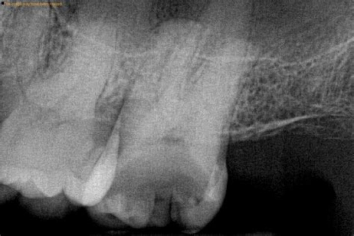
How does a cavity look on an X-ray?
In an X-ray, cavities are seen as dark areas in a tooth. Cavities start at the outside layer covering the tooth, called the Enamel, which has the lightest color in an X-ray. Cavities will then advance to the layer under enamel, called the Dentin, which is softer and has a darker color than enamel in an X-ray.
What are the three types of dental images?
There are three types of diagnostic radiographs taken in today’s dental offices — periapical (also known as intraoral or wall-mounted), panoramic, and cephalometric. Periapical radiographs are probably the most familiar, with images of a few teeth at a time captured on small film cards inserted in the mouth.
What does dental nerve pain feel like?
Tooth nerve pain can feel like a sharp, stabbing pain or a dull ache. If your tooth nerve is exposed, particular foods and drinks will probably trigger the pain.
What does an infected tooth nerve feel like?
In short, when you need a root canal, it may feel like throbbing pain due to infection inside of the root of your tooth. A visible fistula, swelling, or temperature sensitivity might be present. Bacteria can also lead to foul-tasting drainage along the gum tissue near your root.
What does a dying tooth nerve feel like?
The pressure of swollen blood vessels on the pulp nerves will cause pain that could signal to you that you might have a dead tooth. This signal often comes in the form of spontaneous pain, pain when biting or chewing, or extreme sensitivity when drinking hot or cold beverages.
What does a cavity look like in a tooth?
What Does a Cavity Look Like? While it is usually difficult to see a cavity in its beginning stages, some cavities start with a whitish or chalky appearance on the enamel of your tooth. More serious cases can have a discolored brown or black color. However, most often there are no distinguishable red alerts.
What do cavities look like on a dental X-ray?
In an X-ray, cavities are seen as dark areas in a tooth. Cavities start at the outside layer covering the tooth, called the Enamel, which has the lightest color in an X-ray. Cavities will then advance to the layer under enamel, called the Dentin, which is softer and has a darker color than enamel in an X-ray.
What does tooth decay Cry with?
Tooth decay is crying with dental mirror. And sugar coated chocolate on the side. On a white background African American With Sore Tooth Decay. Dental Oral Health Tooth Decay On X-Ray. Tooth Decay On Teeth X-Ray Pain Of Tooth Decay On X-Ray. Pain Of Tooth Decay On Teeth X-Ray Tooth decay is crying with emotions sugar coated chocolate. On the side.
Why do dentists take X-rays of teeth?
Dental x-rays are a diagnostic tool that dentists use to detect conditions and pathology that are not visible during a visual examination. Dental conditions that often require dental x-rays for definitive diagnosis include. Tooth development. Tooth decay between teeth. Tooth abscesses.
What does a periapical dental Xray show?
Periapical (PA) dental x-ray A periapical dental x-ray is a macro image of a single or multiple teeth. The image shows the tooth’s roots and surrounding bone structures. This x-ray enables your dentist to check for the presence of a dental abscess, tissue and/or bone pathology like tumors and cysts and for gum disease.