Inability to follow and object in direction of CN III (the quickest test is to observe upward gaze which is all CN III; the eye on the affected side does not look upward) Inability to open the eyelid. CN III dysfunction causes the eyelid on the affected side to become “droopy”. This is called ptsosis.
Which nerve is tested by the H pattern?
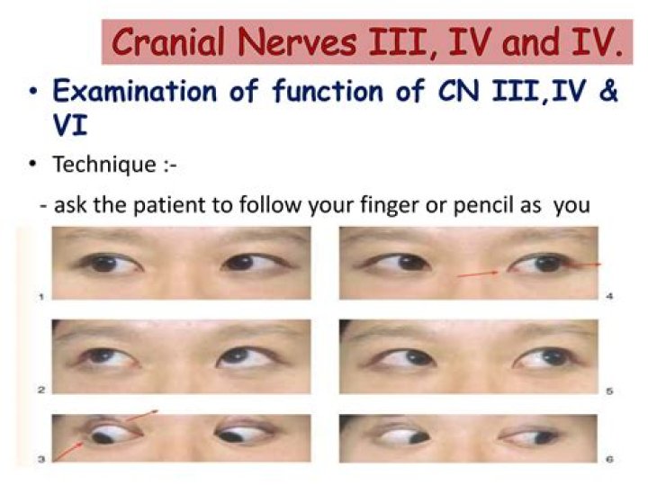
Cranial Nerve 3
Cranial Nerve 3 (Oculomotor):Extraocular muscle testing in “H-pattern” – CN III controls the medial rectus (adduction in to nose), superior rectus, inferior rectus, and inferior oblique. It also controls the elevation of the eyelid, so check for evidence of ptosis.
How is 6th nerve palsy diagnosed?
How to diagnose sixth nerve palsy?
- brain scan to check for a brain tumor, skull fracture, brain injury, or increased pressure in the brain.
- blood test or a lumbar puncture to diagnose or rule out meningitis.
- neurological tests to check for abnormalities in your nervous system.
What type of nerve is the Abducens?
Cranial nerve six (CN VI), also known as the abducens nerve, is one of the nerves responsible for the extraocular motor functions of the eye, along with the oculomotor nerve (CN III) and the trochlear nerve (CN IV).
What does abducens nerve do?
The abducens nerve functions to innervate the ipsilateral lateral rectus muscle and partially innervate the contralateral medial rectus muscle (at the level of the nucleus – via the medial longitudinal fasciculus).
Does the abducens nerve Decussate?
The abducens nerve (or abducent nerve) is the sixth cranial nerve (CNVI), in humans, that controls the movement of the lateral rectus muscle, responsible for outward gaze. It is a somatic efferent nerve….
| Abducens nerve | |
|---|---|
| From | abducens nucleus |
| Innervates | lateral rectus muscle |
| Identifiers | |
| Latin | nervus abducens |
What is abducens nerve palsy?
Sixth nerve palsy occurs when the sixth cranial nerve is damaged or doesn’t work right. It’s also known as the abducens nerve. This condition causes problems with eye movement. The sixth cranial nerve sends signals to your lateral rectus muscle. This is a small muscle that attaches to the outer side of your eye.
What does the abducens nerve do?
What does the abducens nerve pass through?
The abducens nerve passes through the common tendonous ring of the four rectus muscles and then enters the deep surface of the lateral rectus muscle. The function of the abducens nerve is to contract the lateral rectus which results in abduction of the eye. The abducens nerve in humans is solely and somatomotor nerve.
What does Abducens mean?
Definition of abducens nerve : either of the sixth pair of cranial nerves that are motor nerves supplying the rectus on the outer and lateral side of each eye. — called also abducens.
What are the side effects of nerve conduction test?
The nerve conduction study is generally a safe test. However, there are some minor risks associated with this test: You can get an infection where the needle was inserted. Your doctor will talk with you about steps you can take to prevent infection. You may have bleeding at the place where the needle was inserted.
What muscle is innervated by the abducens nerve?
The abducens nerve provides innervation to the lateral rectus muscle – one of the extraocular muscles. The lateral rectus originates from the lateral part of the common tendinous ring, and attaches to the anterolateral aspect of the sclera. It acts to abduct the eyeball (i.e. to rotate the gaze away from the midline).
How do I interpret my EMG results?
The EMG results are generally based around the normal range of the specific muscle being tested so, without knowing what is normal for that area of the body, deciphering the results on your own can be almost impossible. The results of your EMG test should be discussed with a neurologist.
What diseases can be detected by an EMG test?
Amyotrophic Lateral Sclerosis. Amyotrophic lateral sclerosis—ALS,or Lou Gehrig’s disease—is a progressive neuromuscular disease.