An image is formed on the retina with light rays converging most at the cornea and upon entering and exiting the lens. Rays from the top and bottom of the object are traced and produce an inverted real image on the retina. The distance to the object is drawn smaller than scale.
What is the white part of the eye called?
sclera
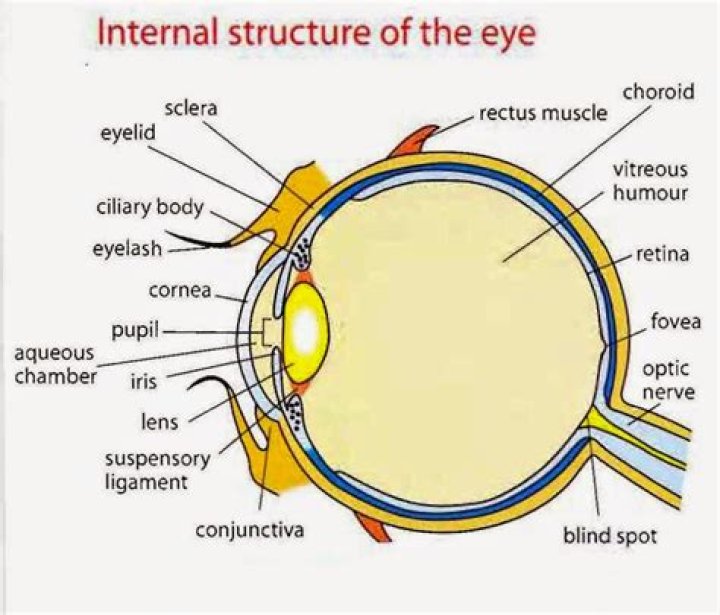
The outer layer of the eyeball is a tough, white, opaque membrane called the sclera (the white of the eye). The slight bulge in the sclera at the front of the eye is a clear, thin, dome-shaped tissue called the cornea. The middle layer is the choroid.
What is the eye structure?
The eye is made up of three coats, which enclose the optically clear aqueous humour, lens, and vitreous body. The outermost coat consists of the cornea and the sclera; the middle coat contains the main blood supply to the eye and consists, from the back forward, of the choroid, the ciliary body, and the iris.
What color are my eyes?
The color of your eyes depends on how much of the pigment melanin you have in your iris—the colored part of your eyes. The more pigment you have, the darker your eyes will be. Blue, grey, and green eyes are lighter because they have less melanin in the iris. Most people in the world will end up with brown eyes.
How does iridology work?
The technique of iridology is based on the belief that each organ in the human body has a corresponding region in the iris. The results are then compared with an iris chart, which helps in correlating the various parts of the human body with different zones in the iris.
What type of image is formed by human eye?
The type of image is formed in the human eye is real and inverted. Our eye lens produces images on our retina that are similar to those produced by a convex lens that is the converging images.
What is bottom of eye called?
This is a hard protective layer which covers all the eyeball except the cornea. The next layer beneath the sclera, between the retina and the sclera, is called the choroid. It circulates through the front part of the eye and then drains away through an area called the trabecular meshwork, near the base of the iris.
What is human eye?
The human eye is a sense organ that reacts to light and allows vision. Rod and cone cells in the retina are photoreceptive cells which are able to detect visible light and convey this information to the brain. The eye is part of the sensory nervous system.
What definition is the human eye?
The human eye is an organ that reacts to light in many circumstances. As a conscious sense organ the human eye allows vision; rod and cone cells in the retina allow conscious light perception and vision, including color differentiation and the perception of depth. The human eye can distinguish about 10 million colors.
How many stock photos of the human eye are there?
Browse 138,391 human eye stock photos and images available or search for human eye close up or human eye anatomy to find more great stock photos and pictures.
What is the size of the human eye in centimeters?
The typical adult eye has an anterior to posterior diameter of 24 millimetres, and a volume of six cubic centimetres (0.4 cu. in.). The eyeball grows rapidly, increasing from about 16–17 millimetres (about 0.65 inch) at birth to 22.5–23 mm (approx. 0.89 in) by three years of age. By age 12, the eye attains its full size. Components
How many human eye photos are available royalty-free?
1,057,886 human eye stock photos, vectors, and illustrations are available royalty-free.
What is the normal field of view of the human eye?
The approximate field of view of an individual human eye (measured from the fixation point, i.e., the point at which one’s gaze is directed) varies by facial anatomy, but is typically 30° superior (up, limited by the brow), 45° nasal (limited by the nose), 70° inferior (down), and 100° temporal (towards the temple).