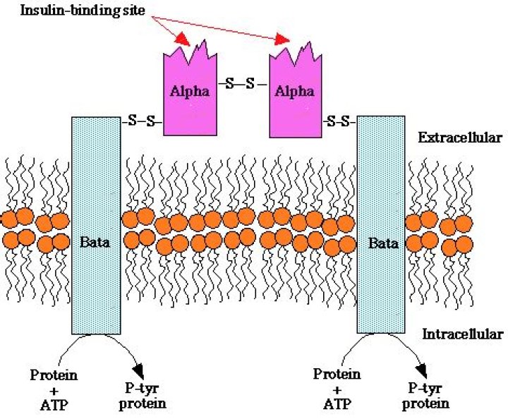
Insulin receptor (IR) is a heterotetramer composed of two extracellular α-subunits and two transmembrane β-subunits, bound together by disulfide bonds.
What type of ligand is insulin?
The insulin receptor is a member of the ligand-activated receptor and tyrosine kinase family of transmembrane signaling proteins that collectively are fundamentally important regulators of cell differentiation, growth, and metabolism.
What receptors receive insulin?
The insulin receptor (IR) is a transmembrane receptor that is activated by insulin, IGF-I, IGF-II and belongs to the large class of receptor tyrosine kinase….Insulin receptor.
| INSR | ||
|---|---|---|
| Species | Human | Mouse |
| Entrez | 3643 | 16337 |
| Ensembl | ENSG00000171105 | ENSMUSG00000005534 |
| UniProt | P06213 | P15208 |
What is the receptor for the insulin hormone?
The receptor belongs to the receptor tyrosine kinase superfamily and has orthologues in all metazoans. The structure of the unbound extracellular domain (“apo-receptor”) has been solved. Insulin binds to two distinct sites on each a subunit of the receptor, crosslinking the two receptor halves to create high affinity.
Where does synthesis of insulin begin?
Insulin is synthesized in significant quantities only in beta cells in the pancreas. The insulin mRNA is translated as a single chain precursor called preproinsulin, and removal of its signal peptide during insertion into the endoplasmic reticulum generates proinsulin.
Is insulin intracellular or extracellular?
The insulin receptor is composed of two alpha subunits and two beta subunits linked by disulfide bonds. The alpha chains are entirely extracellular and house insulin binding domains, while the linked beta chains penetrate through the plasma membrane.
How does insulin act as a ligand?
The α-subunits act as insulin receptors and the insulin molecule acts as a ligand. Together, they form a receptor-ligand complex. Binding of insulin to the α-subunit results in a conformational change in the membrane-bound glycoprotein, which activates tyrosine kinase domains on each β-subunit.
How does insulin work receptor?
Insulin Receptors are areas on the outer part of a cell that allow the cell to join or bind with insulin that is in the blood. When the cell and insulin bind together, the cell can take glucose (sugar) from the blood and use it for energy.
How is insulin receptors made?
The receptor for insulin is a large protein that binds to insulin and passes its message into the cell. It has several functional parts. Two copies of the protein chains come together on the outside of the cell to form the receptor site that binds to insulin.
What does an insulin receptor do?
Where are the α and β subunits of insulin monomers located?
Each monomer is encoded by one gene that forms a single-chain proreceptor that is proteolytically cleaved at a furin cleavage site to form the α and β subunits. The α subunits are located extracellularly and are the location of insulin binding.
Is the insulin receptor extracellular or intracellular?
The α subunit of the insulin receptor is entirely extracellular and contains the insulin-binding domain. The β subunit has an extracellular domain, a transcellular domain, and an intracellular domain that express insulin-stimulated kinase activity directed toward its own tyrosine residues.
How does mutagenesis affect the insulin receptor?
Insulin Receptor/Insulin Receptor Tyrosine Kinase. Similarly, mutagenesis of any of the three major phosphorylation sites (at residues 1158, 1163, and 1162) impairs the insulin receptor kinase activity, and this is associated with a marked decrease in the acute metabolic and growth-promoting effects of insulin.
What is the difference between EX11 and EX11+ insulin receptors?
The insulin receptor exists in two isoforms differing by the absence (Ex11-; IR type A) or presence (Ex11+; IR type B) of 12 amino acids in the C-terminus of the alpha-subunit due to alternative splicing of exon 11. Ex11-binds insulin with two-fold higher affinity than Ex11+.