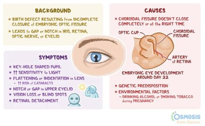
Coloboma arises from abnormal development of the eye. During the second month of development before birth, a seam called the optic fissure (also known as the choroidal fissure or embryonic fissure) closes to form the structures of the eye. When the optic fissure does not close completely, the result is a coloboma.
When does coloboma develop?
Coloboma means that part of one or more structures inside an unborn baby’s eye does not fully develop during pregnancy. This underdeveloped tissue is normally in the lower part of the eye and it can be small or large in size. A coloboma occurs in about 1 in 10,000 births and by the eighth week of pregnancy.
Is there a cure for optic nerve coloboma?
Coloboma of the optic nerve may occur sporadically, may be due to a genetic mutation and be inherited , or may occur as a feature of an underlying syndrome or other genetic condition. There is no treatment to correct an optic nerve coloboma, but low vision aids may be helpful for some people.
How is coloboma diagnosed?
To diagnose a coloboma, an ophthalmologist will perform a comprehensive eye exam using an ophthalmoscope, as well as a visual acuity test. Coloboma of the iris, which gives a “keyhole” appearance of the pupil, does not usually result in vision loss.
Where is coloboma most common?
Eyelid colobomas result in a full-thickness defect of the eyelid: although the coloboma may occur anywhere on the eyelids, the most common site is at the junction of the medial and middle third of the upper eyelid.
How many types of coloboma are there?
Depending on where the closure did not happen, the baby can have an iris coloboma (front of the fissure), a chorio-retinal coloboma (back of the fissure), or any combination of these. Uveal coloboma can affect one eye (unilateral) or both eyes (bilateral).
Can coloboma cause blindness?
Some cases may go unnoticed because uveal coloboma does not always affect vision or the outside appearance of the eye. Uveal coloboma is a significant cause of blindness.
Is there a surgery for coloboma?
We describe a surgical technique for managing congenital iris coloboma. After phacoemulsification with placement of an intraocular lens in the capsular bag, coloboma repair is begun by bisecting the iris sphincter on both sides of its attachment near the chamber angle.
What is complete coloboma?
Coloboma comes from a Greek word which means “curtailed”. It is used to describe conditions where normal tissue in or around the eye is missing from birth.
What is optic disc pit?
Optic disc pit (ODP) is a rare congenital anomaly of the optic disc, which can be complicated by a maculopathy associated with progressive visual loss. Optic disc pits are usually unilateral and sporadic in occurrence, and the development of maculopathy is unpredictable with no known triggers.
What are the symptoms of coloboma?
Keyhole-shaped pupil
How does a coloboma affect vision?
The effect that coloboma has on vision depends on which structures of the eye are involved and how much tissue is missing. Coloboma can affect your iris, the tissue that gives you your eye colour. Your pupil may look oval if the coloboma is partial, but if more of your lower iris is missing, your pupil will look more keyhole shaped.
What are the different kinds of coloboma?
Types of Coloboma Lens Coloboma. A lens coloboma happens when the structures (ciliary body and zonules) maintain the lens in place have a focal defect. Macular Coloboma. The center of the retina is called the macula. Eyelid Coloboma. In cases of eyelid coloboma, the fetus is missing a part of the upper or lower eyelid. Uveal Coloboma. Optic Nerve Coloboma.
What is the structure and function of the optic disc?
The optic disc is where the retinal aqueous and vitreous humor enter and exit the eyeball and where the optic nerve enters and exits the eyeball . The optical disc has no photoreceptors, and therefore marks the blind spot of the eye.