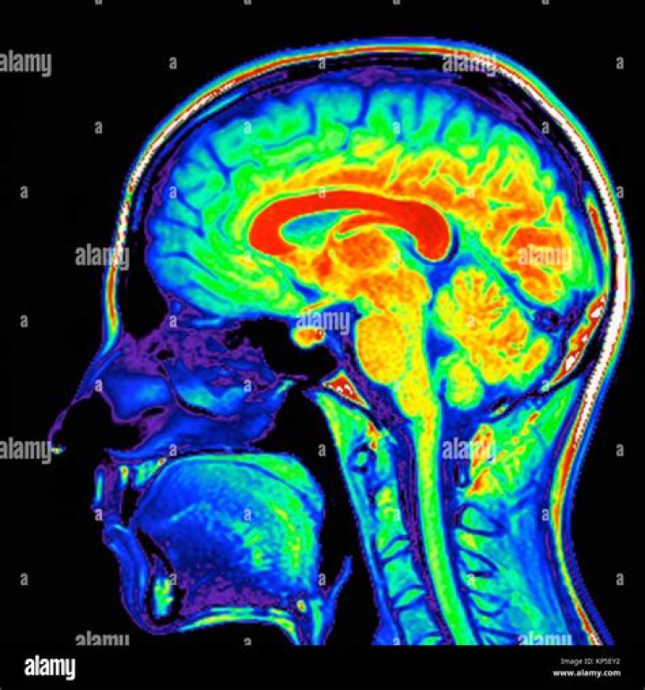
A computer uses the absorption data to show the levels of activity as a color-coded brain map, with one color (usually red) indicating more active brain areas, and another color (usually blue) indicating the less active areas.
What does yellow mean on a brain scan?
Image caption, Daydream Believer: an MRI scan of the brain at rest. Regions in yellow are strongly linked to the area indicated by the blue spot. Image caption, It’s Nice To Be With You: yellow and red areas are involved in processing social interactions.
What is black on a brain MRI?
Air and hard bone do not give an MRI signal so these areas appear black. Bone marrow, spinal fluid, blood and soft tissues vary in intensity from black to white, depending on the amount of fat and water present in each tissue and the machine settings used for the scan.
What lights up on a brain MRI?
On CT or MRI scans, brain lesions appear as dark or light spots that don’t look like normal brain tissue. Usually, a brain lesion is an incidental finding unrelated to the condition or symptom that led to the imaging test in the first place.
What does green mean in a brain scan?
A PET scan can compare a normal brain (left) with one affected by Alzheimer’s disease (right). The loss of red color with an increase in yellow, blue and green colors shows areas of decreased metabolic activity in the brain due to Alzheimer’s disease.
What color is a tumor on MRI?
Dense tumor calcifications are black (signal voids) on MRI, but calcified foci are usually scattered within the soft tissue mass of a tumor, and not liable to be confused with a clear, normal sinus.
What is the colorful brain scan?
This colorful brain scan is a 3D model created by tractography, which uses data collected with diffusion weighted MRI to map the brain’s white matter. Each line represents a bundle of nerve fibers wrapped in a myelin sheaths, which insulate the fibers thereby speeding up the rate of communication between brain regions.
What is white on MRI?
What Are White Spots? Spots on a brain MRI are caused by changes in water content and fluid movement that occur in brain tissue when the brain cells are inflamed or damaged. These lesions are more easily seen on T2 weighted images, a term that describes the frequency (speed) of the radio impulses used during your scan.
What color is blood on MRI?
Oxygenated (arterial) blood is bright red, while dexoygenated (venous) blood is dark reddish-purple. The difference is color results from the electronic state of the iron ion (ferrous vs ferric), which in turn influences the π → π* and n → π* electronic transitions of porphyrin and hence its optical characteristics.
What shows up as white on an MRI?
What brain scan shows colors?
The images of brain PET scans appear as multicolored images of the brain, ranging from dark blue to deep red. Areas of active brain activity come up in warmer colors, such as yellow and red. Your doctor will look at these scans and check for abnormalities.
What does an MRI of the brain show?
Anatomy of the brain (MRI) Cerebellum with its various fissures and lobes as well as the structures of the cerebellar vermis.
How do I use the MRI brain cross sectional anatomy tool?
This MRI brain cross sectional anatomy tool is absolutely free to use. Use the mouse scroll wheel to move the images up and down alternatively use the tiny arrows (>>) on both side of the image to move the images.
How do you describe the colour of tissue in an MRI?
When describing most MRI sequences we refer to the shade of grey of tissues or fluid with the word intensity, leading to the following absolute terms: high signal intensity = white. intermediate signal intensity = grey. low signal intensity = black. Often we refer to the appearance by relative terms:
How long does it take to label 524 structures on MRI?
The elaboration of this new module, its labeling of more than 524 structures on 379 MRI images in three different views and on 26 anatomical diagrams, took more than 6 months.