By ultrasound, the appearance will resemble “Mickey Mouse ears.” Additionally, a measurement from the top of head to the rump (crown rump length or CRL) is significantly reduced in affected fetuses in the first trimester.
How is anencephaly diagnosed?
How is anencephaly diagnosed?
- Alpha-fetoprotein. A protein produced by the fetus that is excreted into the amniotic fluid.
- Amniocentesis. A test performed to determine chromosomal and genetic disorders and certain birth defects.
- Ultrasound (also called sonography).
- Blood tests.
Can anencephaly be diagnosed before birth?
Diagnosis and Tests Providers can also diagnose anencephaly at birth based on the newborn’s appearance. Prenatal tests for anencephaly include: Quad marker screen: This blood test checks for neural tube defects and genetic disorders. Your provider takes a sample of your blood and sends it to a lab for testing.
What is anencephaly ultrasound?
Anencephaly is a lethal congenital anomaly which can be detected on ultrasound as early as 11 weeks of gestation. If some amount of neural tissue is present, the condition is termed exencephaly. Polyhydramnios is usually associated with neural tube defects.
When is anencephaly usually diagnosed?
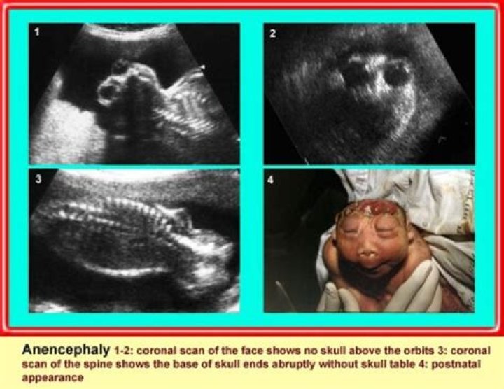
Fetuses with anencephaly are correctly identified at 12 to 13 weeks gestation. Anencephaly occurs in absence of the cranial vault. Ultrasound findings can be normal until onset of ossification has definitely failed. A first trimester scan at 12 to 13 weeks allows reliable diagnosis and active management of anencephaly.
When can you tell a baby has anencephaly?
Tests done during pregnancy to check for anencephaly include: Blood test. A test called a quad screen measures 4 substances in the mother’s blood to see if there is an increased risk for neural tube defects and other problems. This test is done between 16 and 18 weeks of pregnancy.
When is anencephaly diagnosed?
When can you see anencephaly on an ultrasound?
How do you know if your baby has anencephaly?
The diagnosis of anencephaly may be made during pregnancy. Tests done during pregnancy to check for anencephaly include: Blood test. A test called a quad screen measures 4 substances in the mother’s blood to see if there is an increased risk for neural tube defects and other problems.
Can you see anencephaly on an ultrasound?
Ultrasound. Anencephaly can theoretically be diagnosed as early as 8 weeks; however, it can be missed in the first trimester. There is 100% accuracy in the second trimester for this diagnosis by ultrasound.
Is anencephaly a chromosomal abnormality?
Anencephaly can be a multifactorial condition meaning that multiple genes are involved interacting with environmental agents and chance events to cause the condition. Anencephaly can also be a feature of some chromosomal disorders such as trisomy 18 which are usually sporadic and not familial (inherited).
Can you have a baby with anencephaly at 37 weeks?
Most fetuses with anencephaly deliver around 37 weeks of gestation (Melnick and Myrianthopoulos,1987). Because pregnancy with a fetus with anencephaly carries an increased medical risk for the mother, prospective parents may be offered the opportunity to terminate, especially if the diagnosis is made prior to 24 weeks of gestation.
What does anencephaly look like in the first trimester?
In first trimester fetuses with anencephaly, the upper portion of the brain ( the cerebral hemispheres) will be in direct contact with the amniotic fluid because the skull is absent. By ultrasound, the appearance will resemble “Mickey Mouse ears.”
How is brain death diagnosed in babies with anencephaly?
For babies with anencephaly, the diagnosis of brain death depends on documentation of disappearance of previously existing brainstem functions ( i.e., breathing or spontaneous movements ), Most major organs from anencephalic infants are smaller than average for body size and have often not formed appropriately.
Which physical findings are characteristic of anencephaly?
The diagnosis of anencephaly can be confirmed on physical examination when the following criteria are met: a large portion of the skull is absent, the scalp is absent over the skull defect, and a hemorrhagic, fibrotic mass of tissue is exposed to the environment.