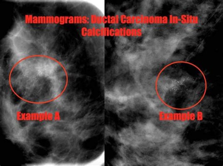
Ductal carcinoma in situ (DCIS) On a mammogram, DCIS usually looks like a cluster of microcalcifications. It can be hard to know from a mammogram whether the cluster is DCIS or invasive breast cancer. A cluster of microcalcifications can also be a benign (not cancer) finding on a mammogram.
Does DCIS show on mammogram?
DCIS is most often discovered during a mammogram used to screen for breast cancer. If your mammogram shows suspicious areas such as bright white specks (microcalcifications) that are in a cluster and have irregular shapes or sizes, your radiologist likely will recommend additional breast imaging.
Does DCIS have a mass?
As mentioned previously, in approximately 2%–23% of cases, DCIS may manifest as a mass or asymmetry at mammography (2,3,5). Noncalcified DCIS may also be detected as a mammographically occult palpable lesion, cause for nipple discharge, abnormality at screening US, or finding in the evaluation of disease extent.
How quickly does DCIS spread?
Grade 1 DCIS is almost always ER and PR positive and is a very slow growing form of cancer. It can take years, even decades, to see progression of the disease. In some cases, it may take such a long time to spread beyond the breast duct that it is not an event that will happen during a person’s lifetime.
What does DCIS lump feel like?
Although DCIS does not usually come with a noticeable lump, the doctor may be able to feel an abnormal growth in the breast, such as a small, hardened spot, during a physical examination. The doctor will also look for any skin changes, nipple changes or nipple discharge.
Can DCIS be missed on a mammogram?
Invasive carcinoma is usually associated with a mass or density found on mammography, whereas DCIS is usually associated with microcalcifications (25). Missed DCIS is much less likely than missed invasive breast cancer to be diagnosed subsequently as a palpable mass, and it may never become clinically apparent.
Where is DCIS located in breast?
Ductal carcinoma in situ (DCIS) is a type of breast cancer. This is also called non-invasive or pre-invasive breast cancer. The cancer cells are found along the sides of the milk duct within the breast.
What is DCIS stage1?
Stage I is the earliest stage of invasive breast cancer. Invasive means that the cancer cells are invading neighboring normal tissue. Stage I breast cancers are 2 centimeters or smaller (a little bigger than 0.75 inches) and have not spread to the lymph nodes.
What does a cancerous tumor look like on a mammogram?
What does breast cancer look like on a mammogram? Any area that does not look like normal tissue is a possible cause for concern. The radiologist will look for areas of white, high-density tissue and note its size, shape, and edges. A lump or tumor will show up as a focused white area on a mammogram.
Is it normal to see lymph nodes on a mammogram?
Normal lymph nodes in the anterior part of the axilla are readily seen on routine mammography. It is important, however, to recognize pathological lymph nodes, characterized by increased attenuation, high density, a round or irregular shape and lack of fat in the hilus, as they often indicate significant diseases.
What causes DCIS breast cancer?
It’s not clear what causes DCIS. DCIS forms when genetic mutations occur in the DNA of breast duct cells. The genetic mutations cause the cells to appear abnormal, but the cells don’t yet have the ability to break out of the breast duct. Researchers don’t know exactly what triggers the abnormal cell growth that leads to DCIS.
What is the prognosis of DCIS breast cancer?
Ductal Carcinoma In Situ (DCIS) Most recurrences happen within the 5 to 10 years after initial diagnosis. The chances of a recurrence are under 30%. Women who have breast-conserving surgery (lumpectomy) for DCIS without radiation therapy have about a 25% to 30% chance of having a recurrence at some point in the future.
Is DCIS cancer or precancer?
DCIS is also called intraductal carcinoma or stage 0 breast cancer. DCIS is a non-invasive or pre-invasive breast cancer. This means the cells that line the ducts have changed to cancer cells but they have not spread through the walls of the ducts into the nearby breast tissue.