The classic radiographic appearance of multiple myeloma is that of multiple, small, well-circumscribed, lytic, punched-out, round lesions within the skull, spine, and pelvis. The pattern of lytic or punched-out radiolucent lesions on the skull have been described as resembling raindrops hitting a surface and splashing.
Can you see multiple myeloma on X ray?
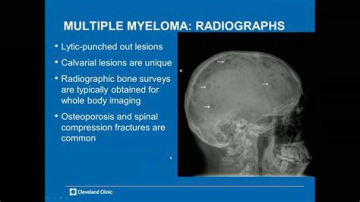
X-rays can detect bone destruction caused by the myeloma cells. Often doctors will do a series of x-rays that includes most of the bones. This is called a bone survey or skeletal survey.
Can multiple myeloma cause shoulder pain?
I previously had researched and found 25% or myeloma patients experience shoulder pain. This is in addition to the pain from bone damage which many myeloma patients already experience.
What are the markers for multiple myeloma?
47·1317 The antigens CD23, CD24, CD25, CD37, CD39, CDw40, and CD45R have also been found in myeloma cells. 7 T-cell antigens (CD2 and CD4) and the neural cell adhesion mol- ecule CD56, a marker for natural killer (NK) cells, may also be present in myeloma cells.
Can myeloma be seen on CT scan?
CT is a sensitive imaging modality in detecting the osteolytic effects of multiple myeloma and has a higher sensitivity than plain radiography at detecting small lytic lesions [16].
What causes pain in shoulder blade?
The most common cause of shoulder blade pain is a muscle strain. 2 Short-term overuse of your arms and upper torso may be experienced in your scapula. This pain may be accompanied by pain in other muscle groups, such as your shoulder or back, but can be felt only in your shoulder blade as well.
Is itching a symptom of multiple myeloma?
This can lead to symptoms such as: Weakness. Shortness of breath. Itching.
Does multiple myeloma show up in a blood test?
Blood work might reveal the abnormal cells that myeloma produces, including M proteins and beta-2-microglobulin. The type of proteins found in the blood can also confirm the aggressiveness of the myeloma. In addition, doctors can use blood work to check for blood cell counts, kidney function, and calcium levels.
Can an X ray of the spine show multiple myeloma?
X-Ray is good for seeing damage in the long bones, but can’t show enough detail in the spine and pelvis Bone damage has to be extensive for it to show up on the X-Ray (30 to 50% bone destruction) It was the gold standard for diagnosing myeloma, but better tools are now available
What imaging studies are used to diagnose multiple myeloma (MM)?
Imaging studies that assess the status of a patient’s bones and/or bone marrow at diagnosis and relapse are: You can learn more about each of these imaging studies below: X-rays are the oldest and least sensitive method to detect myeloma-caused bone damage.
What does multiple myeloma look like on a bone scan?
Bone scintigraphy The bone scan appearance of patients with disseminated multiple myeloma is variable due to the potential lack of osteoblastic activity. Larger lesions may be either hyperactive (hot) or photopenic (cold). Bone scans may also be normal.
What are the symptoms of multiple myeloma with clavicular involvement?
Multiple myelomas may present with shoulder pain and clavicular involvement. The low index of suspicion for myeloma is important as early detection can result in effective treatment and better survival. Uncommon symptoms have been linked to advanced stage myeloma with a reduced life expectancy.