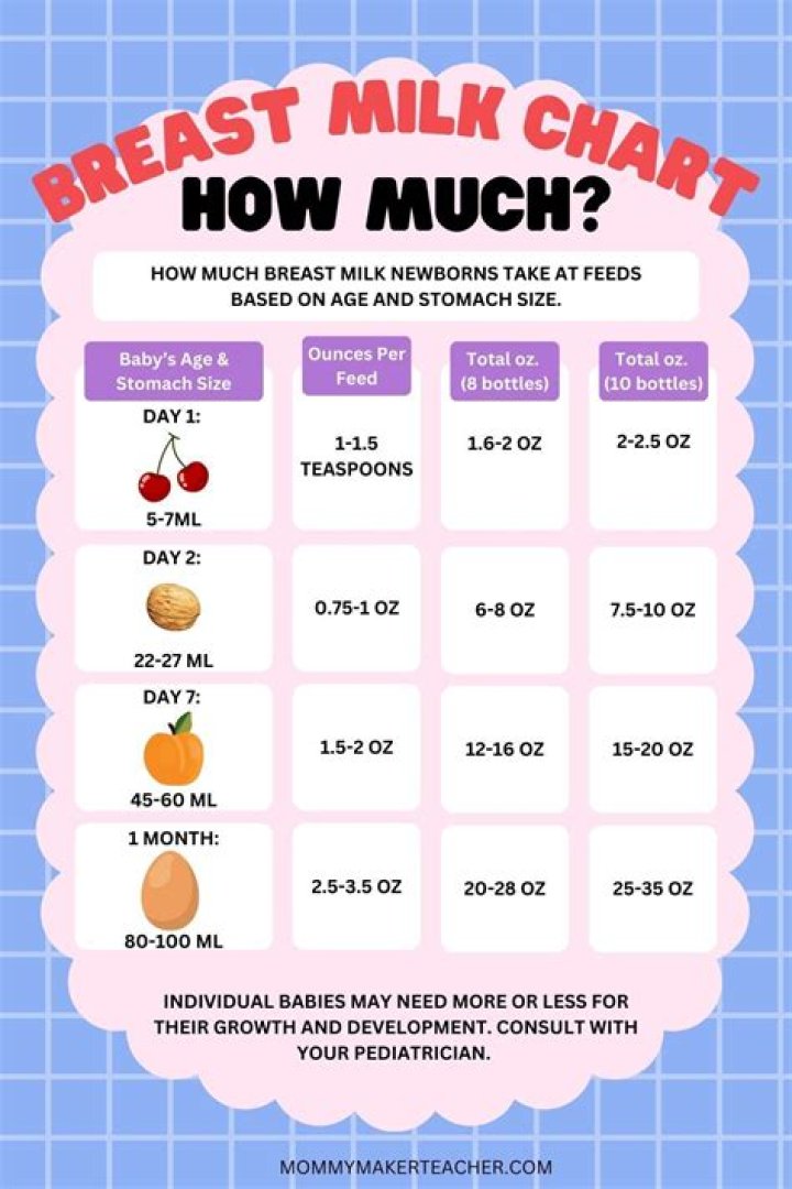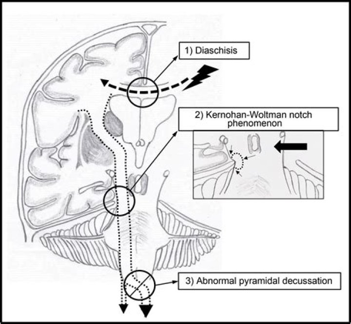
Kernohan notch phenomenon is an imaging finding resulting from extensive midline shift due to mass effect, resulting in the indentation in the contralateral cerebral crus by the tentorium cerebelli. This has also been referred to as Kernohan-Woltman notch phenomenon and false localizing sign.
Why does uncal herniation cause contralateral hemiparesis?
The mass lesion causing the uncal herniation usually causes a contralateral hemiparesis, but as the pressure increases, the opposite cerebral peduncle is compressed against the tentorium, which causes an ipsilateral hemiparesis (Kernohan’s sign).
What is Transtentorial herniation?
A transtentorial herniation is the movement of brain tissue from one intracranial compartment to another. This includes uncal, central, and upward herniation. These are life-threatening and time-critical pathologies that may be reversible with emergent surgical intervention and medical management.
What makes up the Uncus?
The uncus is an anterior extremity of the parahippocampal gyrus. It is separated from the apex of the temporal lobe by a slight fissure called the incisura temporalis. The term comes from the Latin word uncus, meaning hook, and it was coined by Félix Vicq-d’Azyr (1748–1794).
What is an uncal herniation?
Uncal herniation occurs when rising intracranial pressure causes portions of the brain to move from one intracranial compartment to another. It is a life-threatening neurological emergency and indicates the failure of all adaptive mechanisms for intracranial compliance.
What is true about crus cerebri?
The cerebral crus (crus cerebri) is the anterior portion of the cerebral peduncle which contains the motor tracts, travelling from the cerebral cortex to the pons and spine. The plural of which is cerebral crura.
What is ipsilateral pupil dilation?
A unilateral, ipsilateral (on the same side as the lesion), fixed dilated pupil is the initial focal sign, followed by bilateralfixed dilated pupils, occurring anything from minutes to hours later.
How do you declare brain death?
For a diagnosis of brain death:
- a person must be unconscious and fail to respond to outside stimulation.
- a person’s heartbeat and breathing can only be maintained using a ventilator.
- there must be clear evidence that serious brain damage has occurred and it cannot be cured.
Is the amygdala in the uncus?
Abstract. The uncus is the innermost part of the temporal lobe and receives its name from its hook-shaped structure. Anatomically, the anterior segment of the uncus overlies the amygdala and belongs to the parahippocampal gyrus.
What is the Supratentorial?
The supratentorial area (the upper part of the brain) contains the cerebrum, lateral ventricle and third ventricle (with cerebrospinal fluid shown in blue), choroid plexus, pineal gland, hypothalamus, pituitary gland, and optic nerve. The skull and meninges protect the brain and spinal cord (left panel).
What is Kernohan notch phenomenon and what causes it?
Kernohan notch phenomenon is an imaging finding resulting from extensive midline shift due to mass effect, resulting in indentation in the contralateral cerebral crus by the tentorium cerebelli.
What is the Kernohan-Woltman notch phenomenon of ipsilateral motor deficit?
The phenomenon was described by James Watson Kernohan (1896-1981), an Irish-born American pathologist in 1929 after an autopsy study revealed a notched cerebral peduncle from a contralateral herniation syndrome. 1. Zafonte RD, Lee CY. Kernohan-Woltman notch phenomenon: an unusual cause of ipsilateral motor deficit.
How does trans-tentorial herniation cause Kernohan’s notch?
Thus, if you have a right hemisphere trans-tentorial herniation, it causes a Kernohan’s notch in the left cerebral peduncle which results in right-sided motor impairment. Therefore, you get, paradoxically, impairment of motor function on the same side of the body as the herniation which caused the Kernohan’s notch on the controlateral side.
What causes Kernohan’s notch in traumatic brain injury?
Chronic subdural hematomas have been known to be a familiar cause of Kernohan’s notch. MRIs have shown evidence of Kernohan’s notch from patients with traumatic head injury that are related to acute space-occupying lesions such as subdural hematoma, epidural hematoma, depressed skull fracture, or spontaneous intracerebral hematoma.