Normal T axis range between 15o -75o [3]. This is the marker of ventricular repolarization. It is similarly affected by factors affecting the QRS axis. When deviated by >60O in either direction is said to be associated with a cardiac event in adults [4,5].
What does the T-wave of the ECG correlate with?
The T Wave indicates ventricular repolarization, in which the ventricles relax following depolarization and contraction. The ST segment refers to the gap (flat or slightly upcurved line) between the S wave and the T wave, and represents the time between ventricular depolarization and repolarization.
How do you find the T-wave axis?
T-axis was determined by analysing in which frontal leads the highest T-waves were seen. The frontal QRS-T angle was calculated as the absolute value of difference between QRS- and T-axis yielding values between 0 and 180°, and it was categorized as normal (≤90°) or abnormal (≥100°).
What is frontal axis T?
Background and aims: The orientation of the frontal plane T-wave axis (T axis) is a reliable measure of ventricular repolarisation. We investigated the association between T-axis and the risk of coronary heart disease (CHD), heart failure (HF), atrial fibrillation (AF), stroke and cardiovascular (CVD) mortality.
What is a good T wave axis?
The frontal plane T-wave axis was estimated from 12-lead electrocardiograms obtained on admission and categorized as normal (15 degrees to 75 degrees ), borderline (75 degrees to 105 degrees or 15 degrees to -15 degrees ), and abnormal (>105 degrees or < -15 degrees ).
What is an abnormal T wave?
T‐wave abnormalities in the setting of non‐ ST ‐segment elevation acute coronary syndromes are related to the presence of myocardial edema. High specificity of this ECG alteration identifies a change in ischemic myocardium associated with worse outcomes that is potentially reversible.
What does T wave in normal ECG indicate?
Normally, the T wave on an electrocardiogram (ECG) is representative of ventricular repolarization. Changes in T wave morphology can be indicative of various benign or pathologic conditions affecting the myocardium.
What does abnormal T-waves in an ECG mean?
Problem/Condition. The electrocardiographic T wave represents ventricular repolarization. Abnormalities of the T wave are associated with a broad differential diagnosis and can be associated with life-threatening disease or provide clues to an otherwise obscure illness.
What causes T-wave inversion on an ECG?
Inverted T waves. Ischemia: Myocardial ischemia is a common cause of inverted T waves. Inverted T waves are less specific than ST segment depression for ischemia, and do not in and of themselves convey a poor prognosis (as compared to patients with an acute coronary syndrome and ST segment depression).
What causes an abnormal T-wave reading?
Primary T-wave abnormalities (ischemia or injury) are due to alterations in myocardial cellular electrophysiology and secondary T-wave abnormalities (bundle branch block or ventricular Hypertrophy) are subsequent to alterations of sequence of ventricular activation.
What is a good T-wave axis?
What is an abnormal T-wave?
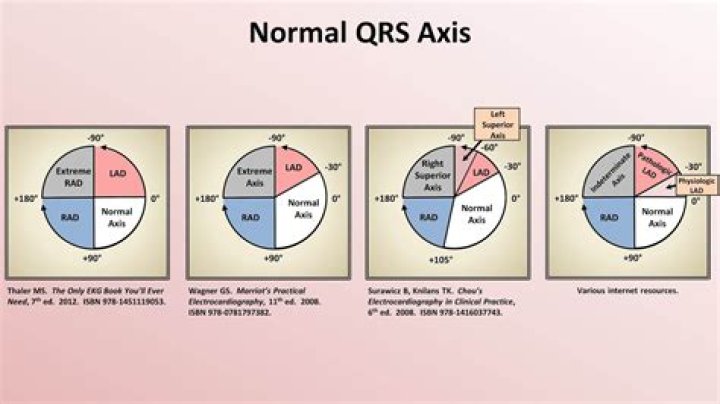
How to calculate ECG axis?
– Identify the most isoelectric lead on the ECG and then on the hexaxial reference diagram – Find the axis line that crosses this lead at 90° – Determine the direction of the axis line by via the lead trace on the ECG.
What is a normal T wave axis?
The frontal plane T-wave axis was estimated from 12-lead electrocardiograms obtained on admission and categorized as normal (15 degrees to 75 degrees ), borderline (75 degrees to 105 degrees or 15 degrees to -15 degrees ), and abnormal (>105 degrees or < -15 degrees ).
What is the T wave axis?
The T-wave axis shift has been reported to represent a general marker of ventricular repolarization abnormalities and a potential indicator of increased risk for cardiovascular mortality.
What is the normal reading for an ECG?
Normal heart rhythm. Overview. An electrocardiogram (ECG) test measures the electrical activity of the heart. A normal resting heart rate is 60 to 100 beats per minute.