The rash typically starts as a red inflammatory papule that expands in a circular or oval shape over several days or weeks to an average size of 16 cm. Central clearing occurs simultaneously. At times, the primary papule or proximal redness remains, contributing to the “bulls-eye” or “target” appearance.
What do you call a ring-like lesion with central clearing?
Annular lesions are rings with central clearing. Examples include granuloma annulare Granuloma Annulare Granuloma annulare is a benign, chronic, idiopathic condition characterized by papules or nodules that spread peripherally to form a ring around normal or slightly depressed skin.
What is annular lesion?
DEFINITION. Annular skin lesions are figurate lesions characterized by a ring-like morphology. Although plaques represent the most common presentation of annular lesions, lesions may also be macular, nodular, or composed of grouped papules, vesicles, or pustules.
Do hives have central clearing?
Urticaria (hives) are raised, erythematous, well-demarcated pruritic skin lesions. Central clearing can cause an annular lesion and is often seen after antihistamine use.
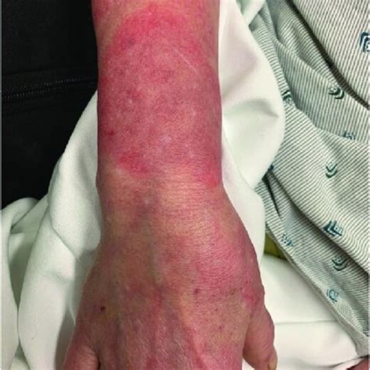
Can eczema clear centrally?
Nummular Eczema (See Figure 9.) Acutely, lesions may ooze and have associated vesicles. Most will lack central clearing, although in some cases, plaques can expand and clear centrally, conferring an annular appearance.
What is the significance of the lesion red border?
A mixture of red and white lesions suggests an irregular epithelial surface that may be caused by a variety of processes, including chronic trauma, inflammation and neoplasia.
What causes EAC?
The cause of EAC is unknown but it is probably due to a hypersensitivity reaction to a variety of agents including drugs, insect or spider bites, infections (bacterial, mycobacterial, viral, fungal), food ingestion (such as blue cheese), malignancies or some underlying diseases.
How do you get rid of an annular lesion?
Annular Lichen Planus of the Glans Penis. Cutaneous LP often is self-limiting; most cases resolve spontaneously within one year. For cutaneous disease, topical corticosteroids under occlusion can be used. When lesions are symptomatic or when oral lesions are present, intralesional triamcinolone is helpful.
What is a polycyclic lesion?
Polycyclic lesions present as configurations arranged in more than one ring. Some annular lesions may coalesce into a polycyclic shape; alternatively, the appearance of such lesions may be independent. For example, the annular lesions of tinea corporis can combine to create a polycyclic lesion.
What is the difference between urticaria and erythema?
While the lesions associated with erythema multiforme, serum-sickness-like reactions, or urticarial vasculitis are fixed and last days to weeks, the skin changes of urticaria multiforme are transient and last less than 24 hours, similar to acute urticaria or juvenile idiopathic arthritis (Still’s disease).
What is the difference between a papule and a target lesion?
Typically, a papule or plaque expands with erythematous borders, while the center may become necrotic or dusky, resulting in a “target lesion.”. Psoriasis, which most commonly presents as erythematous plaques with diffuse thick, white scale, can present as annular lesions with scale only on the borders (Figure 8).
How are annular lesions diagnosed and treated?
Annular Lesions: Diagnosis and Treatment Annular lesions can present in a variety of diseases. Knowledge of the physical appearance and history of presentation of these skin findings can help in the diagnosis. A pruritic, annular, erythematous patch that grows centrifugally should prompt evaluation for tinea corporis.
What are perforating and subcutaneous granuloma annulare lesions?
Perforating granuloma annulare lesions are small, umbilicated papules that are found predominantly on the hands and fingers. Subcutaneous granuloma annulare is characterized by large, skin-colored nodules that may be as deep as the lower dermis or subcutaneous fat. 1
What is the presentation of subcorneal pustular dermatoses (Sneddon-Wilkinson disease)?
Subcorneal pustular dermatoses (Sneddon-Wilkinson disease) presents as oval, peasized flaccid pustules which rupture easily, and tend to coalesce, forming annular or serpiginous patterns with a scaly edge. Characteristically, pus accumulates in the lower half of a fully developed pustule, leaving clear fluid in the upper half.