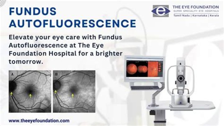
Fundus autofluorescence (FAF) is a non-invasive imaging technique that detects fluorophores, naturally occurring molecules that absorb and emit light of specified wavelengths [1].
How is fundus autofluorescence done?
Fundus autofluorescence (FAF) is a relatively new, non-invasive imaging modality that has been developed over the past decade. The FAF images are obtained through the use of confocal laser scanning ophthalmoscopy (cSLO).
What is hydroxychloroquine eye test?
“Today it is recommended that every single person on Plaquenil get a visual field 10-2 test, plus one of the three other highly sensitive screening tests: the FAF (fundus autofluorescence imaging), the SD-OCT (spectral domain optical coherence), or the multifocal electroretinogram (mfERG),” Thomas says.
How does hydroxychloroquine affect the retina?
Plaquenil binds to retinal pigment cells, causing adverse changes in vision that occur slowly over time. Objects may start to appear blurry or distorted. In the most serious cases of Plaquenil-induced retinal toxicity, the macula is completely destroyed.
Why do you need an eye exam with hydroxychloroquine?
Protect Your Vision—Get a Plaquenil Screening One such drug is Plaquenil. Using this medication for a long time can cause damage to the retina, which may lead to vision loss. If you take Plaquenil, call Rock Hill Eye Center to schedule a screening.
Can hydroxychloroquine cause blindness?
It’s generally safe at normal doses, but higher amounts can damage the retina, the light-sensitive tissue at the back of the eye, and result in partial or complete blindness.
What are the serious side effects of hydroxychloroquine?
Hydroxychloroquine may cause side effects. Tell your doctor if any of these symptoms are severe or do not go away:
- headache.
- dizziness.
- loss of appetite.
- nausea.
- diarrhea.
- stomach pain.
- vomiting.
- rash.
What kind of eye problems does hydroxychloroquine cause?
It is known that some people who take hydroxychloroquine for more than five years and/or in high doses are at increased risk of damage to their retina, the light sensitive layer of cells at the back of the eye. This is known as retinal toxicity or retinopathy.
How do I reduce autofluorescence?
Historically, the main method that has been employed to lower autofluorescence has been to treat the tissue with solutions of Sudan Black or similar dyes. These hydrophobic dye molecules can bind to tissue sections and lower fluorescence through absorption of incident radiation.
What does increased autofluorescence in fundus mean?
Increased autofluorescence is seen corresponding to the vitelliform deposits, along with mottled increased and decreased autofluorescence, which is typical of pattern dystrophies. Fundus autofluorescence may be used to monitor progression of a variety of inherited retinal diseases.
What is FAF undus autofluorescence used for?
F undus autofluorescence (FAF) is a noninvasive tool to characterize the structure and function of the retinal pigment epithelium (RPE)/RPE complex. As an imaging modality, it may often detect abnormalities not seen on clinical examination.
How much Hydroxychloroquine is safe for retinal toxicity?
In summary, hydroxychloroquine-induced retinal toxicity is likely more prevalent than previously believed. Once thought to be safe at a daily dose of <6.5mg/kg/day [16], newer studies show that many years of hydroxychloroquine treatment at a “safe dose” can still lead to toxicity [6].
Is corneal verticillata a sign of hydroxychloroquine toxicity?
The finding of corneal verticillata bares no correlation with retinal toxicity and is not an indication to stop the medication [ 1 ]. The hallmark of hydroxychloroquine toxicity is bilateral pigmentary retinopathy [ 1 ]. Patients with early retinal findings are often asymptomatic despite having subtle paracentral scotomas.