The list of entities associated with a high signal intensity on T1-weighted images is extensive and classically includes fat, proteins, hemorrhage, melanin and gadolinium.
What does high T1 signal mean?
T1 weighted image – Pathology (spine) Loss of the normal high signal in the bone marrow indicates loss of normal fatty tissue and increased water content. Abnormal low signal on T1 images frequently indicates a pathological process such as trauma, infection, or cancer.
What is a T1 weighted MRI image?
Definition. A T1-weighted (T1W) image is a basic pulse sequence in magnetic resonance (MR) imaging and depicts differences in signal based upon intrinsic T1 relaxation time of various tissues.
What is T1 weighted image good for?
T1-weighted sequences provide the best contrast for paramagnetic contrast agents such as gadolinium-containing compounds. These are areas where the disease that are currently active. Before the MRI, an injection of gadolinium (gd) is administered.
What does high signal on MRI mean?
High signal seen on these images indicates a pathological process such as infection, tumour, or areas of demyelination – as in this patient with multiple sclerosis.
What is a high signal lesion?
In the current study, a mass with a lesion/fat signal intensity ratio of greater than 0.7 on a T1-weighted sequence was considered high signal intensity.
What does signal changes on brain MRI mean?
Spots on a brain MRI are caused by changes in water content and fluid movement that occur in brain tissue when the brain cells are inflamed or damaged. These lesions are more easily seen on T2 weighted images, a term that describes the frequency (speed) of the radio impulses used during your scan.
What does T1 shortening mean?
Contrast enhanced The most commonly used contrast agents in MRI are gadolinium based. At the concentrations used, these agents have the effect of causing T1 signal to be increased (this is sometimes confusingly referred to as T1 shortening).
What is T1 signal intensity?
T1 is the time required for spins to recover approximately 63% of their preexcitation magnetization. Substances that have intrinsically shorter T1 relaxation times demonstrate higher signal intensity at T1-weighted imaging.
What are T1 lesions?
T1 lesions were defined as regions with a signal intensity similar to or reduced to the signal intensity of gray matter and corresponding to a hyperintense region on T2-weighted MRI. Hyperintense–T2 lesions were defined as sharply demarcated regions of high signal intensity compared with surrounding brain tissue.
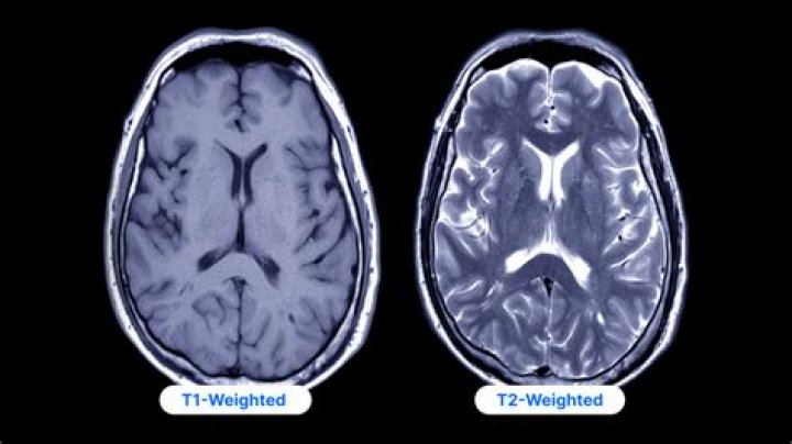
What does T1 and T2 weighted images mean?
T1-weighted images are produced by using short TE and TR times. The contrast and brightness of the image are predominately determined by T1 properties of tissue. Conversely, T2-weighted images are produced by using longer TE and TR times.
How can you tell the difference between T1 and T2 weighted MRI?
The best way to tell the two apart is to look at the grey-white matter. T1 sequences will have grey matter being darker than white matter. T2 weighted sequences, whether fluid attenuated or not, will have white matter being darker than grey matter. Read more about FLAIR sequence.
What is T1 imaging?
The basis of T1 weighted imaging is the longitudinal relaxation. A T1 weighted magnetic resonance image is created typically by using short TE and TR times. The final image is a reflection of more than one of these pulse sequence parameters, weighted according to the type of sequence and its timing.
What is proton T1 weighted MRI?
T1 weighted image (also referred to as T1WI or the “spin-lattice” relaxation time) is one of the basic pulse sequences in MRI and demonstrates differences in the T1 relaxation times of tissues. A T1WI relies upon the longitudinal relaxation of a tissue’s net magnetization vector (NMV).
What are the T1 and T2 signals in a MRI?
One of these, probably T1, is a measure of the tissues’ responses to the signal; the other, probably T2, is the measure of the tissues’ relaxation speed after stimulation by the MRI machine. So, basically, the machine makes a signal, the tissues respond, the device records the response of the tissues to the signal.
What is a MRI image?
A magnetic resonance imaging (MRI) scan is an imaging test that uses powerful magnets and radio waves to create pictures of the body. It does not use ionizing radiation (x-rays). Single MRI images are called slices. The images can be stored on a computer or printed on film.