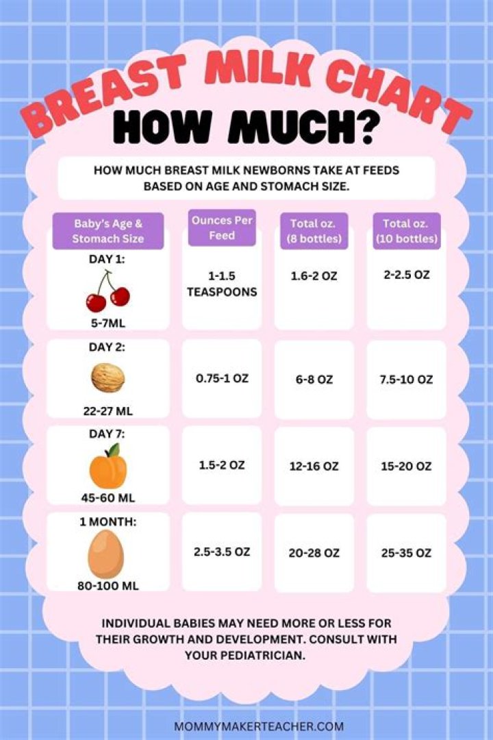
approximately 4:1
There is a normal ratio of myeloid to erythroid precursors (approximately 4:1) with normal maturation of both cell lines.
What is the M E ratio in megaloblastic anemias?
M:E ratio. The ratio of maturing myeloid cells to erythroid cells in the bone marrow, which is normally 3–4:1. Decreased M:E. Haemolytic and megaloblastic anaemias.
Why there is erythroid hyperplasia in megaloblastic anemia?
The bone marrow is hypercellular with erythroid hyperplasia. Erythroid precursors have megaloblastic features being larger than normoblastic cells. In addition, nuclear maturation is immature relative to cytoplasmic maturation. Megaloblastic changes are most prominent in more mature RBC precursors.
What is the myeloid to erythroid ratio in this patient and what does it indicate?
the ratio of myeloid to erythroid precursors in bone marrow; normally it varies from 2:1 to 4:1; an increased ratio is found in infections, chronic myelogenous leukemia, or erythroid hypoplasia; a decreased ratio may mean a depression of leukopoiesis or normoblastic hyperplasia depending on the overall cellularity of …
How do you calculate myeloid erythroid ratio?
The M/E ratio was determined by dividing the total of all the nucleated cells of the granulocytic series by the total of all the nucleated cells of the erythrocytic series.
What would the M E ratio be for an erythroid leukemia?
The normal M:E ratio in adults varies from 1.2:1 to 5:1 myeloid cells to nucleated erythroid cells. An increased M:E ratio (6:1) may be seen in infection, chronic myelogenous leukemia or erythroid hypoplasia.
What is the M E ratio?
The M:E ratio indicates the relative numbers of myeloid lineage cells (all granulocytic and monocytic cells) to nucleated erythroid precursors in marrow. For example, if the M:E ratio is increased, it indicates that there is either myeloid hyperplasia or an erythroid hypoplasia or a combination of both.
What causes Cabot ring bodies?
Cabot rings are thin, threadlike ring- or “figure eight”–shaped red blood cell inclusions, likely remnants from mitotic spindles. They are rarely seen in peripheral blood, and their presence indicates a defect in erythrocyte production, especially in pernicious anemia and lead poisoning.
What is megaloblastic erythroid hyperplasia?
Definition. A laboratory test result indicating an abnormally high quantity of abnormal immature red blood cells with megaloblastic features. [ from NCI]
What causes erythroid hyperplasia?
Hemorrhage, hemolytic anemia, intrinsic bone marrow disease (including aplastic anemia and malignant neoplasms), and anemia of chronic disease are the most common causes of erythroid hyperplasia associated with normocytic anemia in patients with no history of a toxic insult, chemotherapy, or hemoglobinopathy.
How is myeloid erythroid ratio calculated?
A myeloid-to-erythroid (M : E) ratio (also referred to as a granulocytic-to-erythroid ratio) is calculated by examining 500 cells and dividing the number of granulocytic cells, including mature granulocytes, by the number of nucleated erythroid cells.
How do you solve myeloid erythroid ratio?
What are the causes of megaloblastic anemia?
This anemia is caused due to the deficiency of Vitamin B12 and/or Folic acid. Less commonly, also due to acquired abnormalities of their metabolism. Megaloblastic anemia (MBA) may cause a problem in differential diagnosis from other conditions which may cause macrocytosis.
What is the normal M E ratio for erythropoiesis?
M:E ratio: Due to marked erythroid hyperplasia, M:E ratio is reversed ranging from 1:1 to 1:6 (normal 2:1 to 4:1). Erythropoiesis: Megaloblastic type.
What is the typical ratio of erythroid precursor cells to myeloid precursor cells?
Erythroid hyperplasia, indicated by a reduced ratio of myeloid to erythroid precursor cells (0.7; reference range 2-4:1) and 2% blasts
What is the role of dihydrofolate reductase in megaloblastic anemia?
The reduction of dihydrofolate to tetrahydrofolate by dihydrofolate reductase is targeted by multiple drugs, 5, 13 which have the effect of decreasing available deoxythymidylate for DNA synthesis, resulting in megaloblastic anemia.