0.3 to 1 ug/
The detection of protein bands in the gel by Coomassie Blue staining depends on nonspecific binding of a dye, Coomassie Brilliant Blue R, to proteins. The detection limit is 0.3 to 1 ug/protein band.
What is Coomassie blue r250?
Coomassie R-250 and G-250 dyes are two chemical forms of a disulfonated triphenylmethane compound that is commonly used as the basis of stains for detection of proteins in gel electrophoresis and Bradford-type assay reagents for protein quantitation. …
What is the detection range of SDS PAGE?
It has a detection limit of ~ 0.1–0.5 μg protein, sensitive enough for most daily needs. Silver staining has greater sensitivity, but involves many more steps and solutions (see Silver Staining of SDS-polyacrylamide Gel).
What is the difference between r250 and g250 Coomassie Brilliant Blue G-250?
Basically, both R-250 (R signifies the slightly reddish tint in the blue color of the dye) and G-250 (G signifies the greenish tint in the dye) have relatively high sensitivity and allow for easy detection (they develop intensely colored complexes upon binding with protein molecules).
What is the sensitivity of Coomassie blue?
Sensitivity. 3-10 microgram of protein can be detected usually within 5 minutes in Coomassie stains. With additional water-based de-staining, as little as 7 ng of protein (BSA) can be detected.
Can Coomassie Brilliant Blue interfere in SDS-PAGE?
Having both dyes in a gel is a bad idea. CBB250 binds proteins and changes their mobility in SDS PAGE. The reason why the bromophenol blue dye front is smeared is usually due to high salt in the sample (greater than 0.1M) The BPB dye front should be straight as an arrow if the gel is run properly.
What is the minimum amount of the protein that can be detected by SDS-PAGE method?
Ideally, it is best to load ≤2 µg per well of a purified protein or ≤20 µg of a complex mixture like whole cell lysates if you are doing Coomassie stain only.
Can Coomassie Brilliant Blue interfere in SDS PAGE?
Does SDS-PAGE separate subunits?
In their native form, proteins fold into a variety of shapes, some compact, some elongated. It also separates subunits in multimeric proteins, allowing analysis of large, complex aggregates. The most commonly used denaturant is sodium dodecyl sulfate (SDS).
What is biochemistry page?
Polyacrylamide gel electrophoresis (PAGE) is a technique widely used in biochemistry, forensic chemistry, genetics, molecular biology and biotechnology to separate biological macromolecules, usually proteins or nucleic acids, according to their electrophoretic mobility.
What is the difference between Coomassie R-250 and G-250?
Coomassie R-250, is the most commonly used variant for detection of protein, allowing for detection of as little as 0.1 ug protein. The coomassie G-250 dyes is less sensitive, with a lower limit of around 0.5 µg for most proteins, but this is somewhat offset by the speed of destaining offered by this dye compared to R-250.
What is Coomassie Brilliant Blue R250?
Thermo Scientific Pierce Coomassie Brilliant Blue R-250 is one of the most common forms of coomassie dye, which is a key component of various colorimetric protein gel stains. Coomassie R-250 and G-250 dyes are two chemical forms of a disulfonated triphenylmethane compound that is commonly used as the basis of stains for detection…
How are Coomassie dyes used to quantify proteins?
Laboratory usage. The Coomassie dyes (R-250 and G-250) are used for quantification of protein, and work by binding to proteins through Van der Waals attractions and through ionic interactions between dye sulfonic acid groups and positive protein amine groups . Coomassie R-250, is the most commonly used variant for detection of protein,…
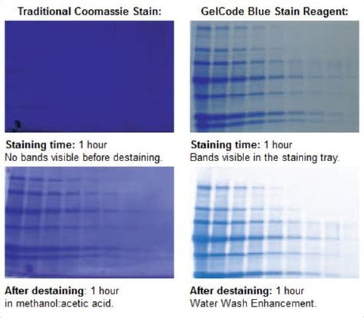
How long does it take to stain with Coomassie Blue R-250?
Stain gel in the above solution, with 0.25% Coomassie Blue R-250, for 2 – 4 hours, until the gel is a uniform blue color. Staining is complete when the gel is no longer visible in the dye solution.