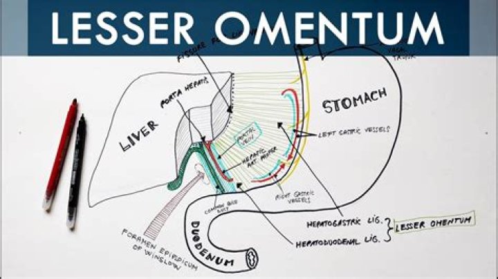
The lesser omentum transports the arteries for the lesser curvature of the stomach; the right and left gastric arteries.
What is found in the lesser omentum?
The free border of the lesser omentum between the porta hepatis and the duodenum contains the hepatic artery, the portal vein, the common bile duct, lymph glands, lymph vessels, and nerves, forming the hepatic hilum. Behind this free edge is the opening into the lesser sac or foramen of Winslow.
What is the omentum quizlet?
A double-layered connecting peritoneum between the stomach and abdominal organs or abdonimal wall. Connects the lesser curvature of the stomach and first part of the duodenum to the porta of the liver. It is continuous aborad (away from the mouth) with the mesoduodenum.
Is lesser sac the same as lesser omentum?
The lesser sac, also known as the omental bursa, is the cavity in the abdomen that is formed by the lesser and greater omentum. Usually found in mammals, it is connected with the greater sac via the omental foramen or Foramen of Winslow….
| Lesser sac | |
|---|---|
| Latin | bursa omentalis |
| TA98 | A10.1.02.402 |
| TA2 | 3703 |
| FMA | 19800 |
What is in the lesser sac?
The lesser omentum is composed of two peritoneal ligaments that extend from the lesser curvature of the stomach and duodenal bulb to the liver, the gastrohepatic, and hepatoduodenal ligaments, respectively.
Where does the lesser omentum attach to?
liver
The lesser omentum goes from the lesser curve here, to the underside of the liver, where its attachment is just out of sight. It’s attached up here to the underside of the diaphragm. The lesser omentum extends down here onto the duodenum, where it has a free lower border as we’ll see.
How many layers of peritoneum are in lesser omentum?
two layers
The lesser omentum is extremely thin, and is continuous with the two layers of peritoneum which cover respectively the antero-superior and postero-inferior surfaces of the stomach and first part of the duodenum.
What is the greater omentum?
The greater omentum is a 4-layered fold of peritoneum that extends down from the stomach, covering much of the colon and small bowel. The layers are generally fused together caudal to the transverse colon. The gastrocolic ligament is part of the greater omentum.
How do the greater and lesser sacs communicate?
The omental bursa or lesser sac is a hollow space that is formed by the greater and lesser omentum and its adjacent organs. It communicates with the greater sac via the epiploic foramen of winslow, which is known as the general cavity of the abdomen that sits within the peritoneum, but outside the lesser sac.
What is the lesser peritoneal sac What are its boundaries?
The lesser sac may be conceptualized as the space posterior to the lesser omentum, between the posterior wall of the stomach and surface of peritoneum that covers the anterior surface of the left kidney 1.
What is the lesser peritoneal sac?
The lesser sac may be conceptualized as the space posterior to the lesser omentum, between the posterior wall of the stomach and surface of peritoneum that covers the anterior surface of the left kidney 1. The epiploic foramen (of Winslow) is the only natural communication between greater and lesser sac.
What does the lesser sac contain?
What is the prognosis for cancer in the omentum?
Cancer of the omentum is linked to ovarian cancer, which generally has a 45 percent survival rate after 5 years, according to the American Cancer Society. In 80 percent of ovarian cancer patients the disease has spread to the omentum before the condition is diagnosed, the University of Chicago Medical Center says.
Is it from the mesentery or the omentum?
The omenta are derived from the embryological ventral and dorsal mesenteries . The greater omentum is derived from the dorsal mesentery, while the lesser omentum originates from the ventral mesentery. The greater omentum (or omentum majus), as its name suggests, is the largest of the two omenta.
What is an omental biopsy?
An omental biopsy involves inserting a needle through the skin to take a sample of the omentum. Local anaesthetic is used to numb the skin. The radiologist uses an ultrasound scan to accurately direct the needle into the area that needs to be sampled.