Eosinophilic describes the appearance of cells and structures seen in histological sections that take up the staining dye eosin. This is a bright-pink dye that stains the cytoplasm of cells, as well as extracellular proteins such as collagen. Such eosinophilic structures are, in general, composed of protein.
Why does eosinophil stain red?
These cells are eosinophilic or “acid-loving” due to their large acidophilic cytoplasmic granules, which show their affinity for acids by their affinity to coal tar dyes: Normally transparent, it is this affinity that causes them to appear brick-red after staining with eosin, a red dye, using the Romanowsky method.
What color should an eosinophil stain with eosin?
The eosin stains proteins (pink). The white blood cells shown here (left panel) are eosinophils (nucleus with 2 lobes) and neutrophils (nucleus with 2-5 lobes). The intense pink staining in the eosinophils is the reason why these cells were named “eosinophils”, meaning “eosin loving”.
How do you identify eosinophils?
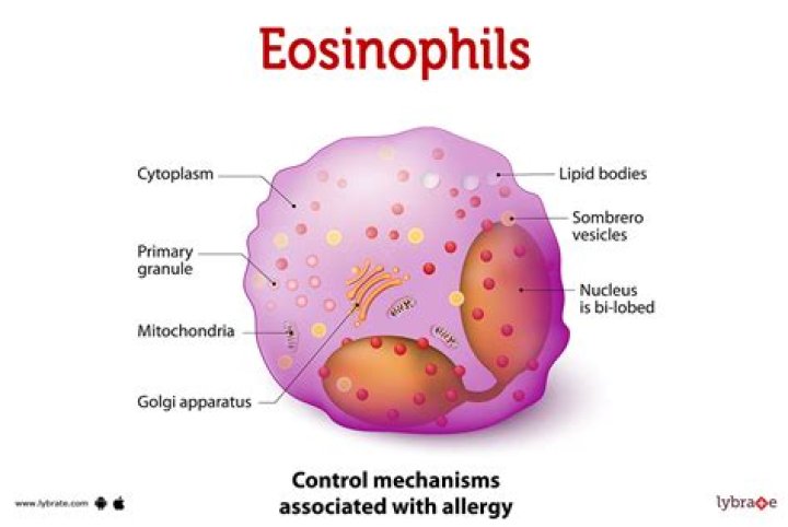
These cells are 12 – 17 µm in diameter – larger than neutrophils, and about 3 times the size of a red blood cell. You can see that eosinophils only have two lobes to their nucleus. These cells have large acidophilic specific granules – these stain bright red, or reddish-purple.
What stains eosinophilic and basophilic?
Eosin is an acidic dye: it is negatively charged (general formula for acidic dyes is: Na+dye-). It stains basic (or acidophilic) structures red or pink. This is also sometimes termed ‘eosinophilic’. It is used to stain acidic (or basophilic) structures a purplish blue.
What is Basophilic staining?
Basophilic describes the appearance of structures seen in histological sections which take up basic dyes. The structures usually stained are those that contain negative charges, such as the phosphate backbone of DNA in the cell nucleus and ribosomes.
What Colour is hematoxylin?
blue-purple
Hematoxylin has a deep blue-purple color and stains nucleic acids by a complex, incompletely understood reaction. Eosin is pink and stains proteins nonspecifically. In a typical tissue, nuclei are stained blue, whereas the cytoplasm and extracellular matrix have varying degrees of pink staining.
What is the Colour of eosinophil?
Eosinophils are generally the largest granulocytes found in normal blood. Their cytoplasm is usually colorless or light blue. However, the color is usually masked by the large granules that are present. These granules take up the acid components of Wright stain, and are therefore reddish-orange.
What is normal range for eosinophils?
Normal eosinophil count is less than 500 cells per microliter (cells/mcL). Normal value ranges may vary slightly among different laboratories. Talk to your provider about the meaning of your specific test results.
Are ribosomes basophilic or eosinophilic?
Most cellular organelles and extracellular matrix are eosinophilic, while the nucleus, rough endoplasmic reticulum, and ribosomes are basophilic.
What are the causes of high eosinophils?
Parasitic and fungal diseases
What medications can cause high eosinophils?
Similarly, food allergies can also cause elevated eosinophil counts. Eosinophilia Esophagitis (EoE): This is a disorder characterized by eosinophils spreading to the esophagus which normally does not contain eosinophils. About 50% of people with EoE will also have elevated eosinophil counts in the blood. 4
What drugs cause eosinophilia?
Aldesleukin
What causes increase in eosinophils?
The most common causes of a high number of eosinophils (called eosinophilia or hypereosinophilia) are Allergic disorders, including drug sensitivities, asthma, allergic rhinitis, and atopic dermatitis, often increase the number of eosinophils.