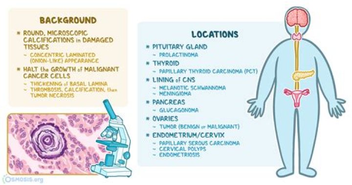
Psammoma bodies can be found in organs such as the thyroid, ovaries, endometrium, and the lining of the central nervous system. They are involved in both cancerous and benign tumors, and can also be a sign of chronic inflammation.
What cancers have Psammoma bodies?
Psammoma bodies are found in a diverse group of tumors which include:
- papillary thyroid carcinoma.
- papillary serous carcinoma of the endometrium.
- melanotic schwannoma (psammomatous variety)
- meningioma.
- mesothelioma.
- serous cystadenocarcinoma of the ovary.
- adenocarcinoma of lung.
What is Psammoma bodies in thyroid?
The presence of psammoma bodies is a diagnostic characteristic of papillary thyroid carcinoma. It is defined as spherical calcified foci with concentric laminations,1, 19, 20 and is usually located within stromal stalks of tumor papillae, and is distinct from intrafollicular inspissated colloid.
Does Prolactinoma have Psammoma body?
Prolactinomas. These tumors are chromophobic and contain spheroid nuclei with prominent nucleolus. Psammoma bodies and interstitial amyloid deposits, which are seen occasionally, are typical for this adenoma type.
When are Psammoma bodies seen?
Psammoma bodies are commonly seen in certain tumors such as: Papillary thyroid carcinoma. Papillary renal cell carcinoma. Ovarian papillary serous cystadenoma and cystadenocarcinoma.
Are Psammoma bodies seen in mesothelioma?
This association of psammoma bodies with a mesothelioma is an exceedingly rare finding. Twenty-five men and 10 women with malignant mesothelioma seen at the Austin Hospital between 1965 and 1978 were reviewed. The patients ranged in age from 34 to 79 yr.
Why Psammoma bodies are formed?
Cause. Psammoma bodies are associated with the papillary (nipple-like) histomorphology and are thought to arise from, Infarction and calcification of papillae tips. Calcification of intralymphatic tumor thrombi.
What is Orphan Annie eye?
Features of Orphan Annie-eye nuclei in histopathology include, large nuclei, oval/molded with singular membranes, nuclear clearing with powdery chromatin, nuclear grooves, one/more marginally placed micro nucleoli, nuclear crowding with optically clear ground glass appearance, and nuclei that are often seen overlapping …
What cancers cause twitching?
Seizures and brain cancer While seizures can be caused by other conditions such as epilepsy, a brain tumor can irritate the neurons in the brain, causing muscle contractions, twitching, numbness and tingling, shallow breathing and loss of consciousness.
What does neoplastic mean?
Listen to pronunciation. (NEE-oh-PLA-zum) An abnormal mass of tissue that forms when cells grow and divide more than they should or do not die when they should. Neoplasms may be benign (not cancer) or malignant (cancer).
Is a Cystadenoma benign or malignant?
Ovarian cystadenomas are common benign epithelial neoplasms which carry an excellent prognosis. The two most frequent types of cystadenomas are serous and mucinous cystadenomas whereas endometrioid and clear cell cystadenomas are rare.
Can psammoma bodies be seen on CT?
They have a lamellated concentric calcified structure, sometimes large enough to be seen on CT. Psammoma bodies are found in a diverse group of tumours which include: papillary thyroid carcinoma. papillary serous carcinoma of the endometrium 2.
What is the pathophysiology of psammoma bodies?
Psammoma bodies are round microscopic calcific collections. It is a form of dystrophic calcification. Necrotic cells form the focus for surrounding calcific deposition.
What are the different types ofpsammoma bodies?
Psammoma bodies are found in a diverse group of tumors which include: 1 papillary thyroid carcinoma. 2 papillary serous carcinoma of the endometrium 2. 3 melanotic schwannoma (psammomatous variety) 3. 4 meningioma. 5 mesothelioma. 6 (more items)
What is psammomatous meningioma?
Dr Mark Thurston ◉ and Dr Saeed Soltany Hosn et al. Psammomatous meningioma is a histologic subtype of meningioma usually presented as a heavily calcified intracranial or spinal mass lesion.