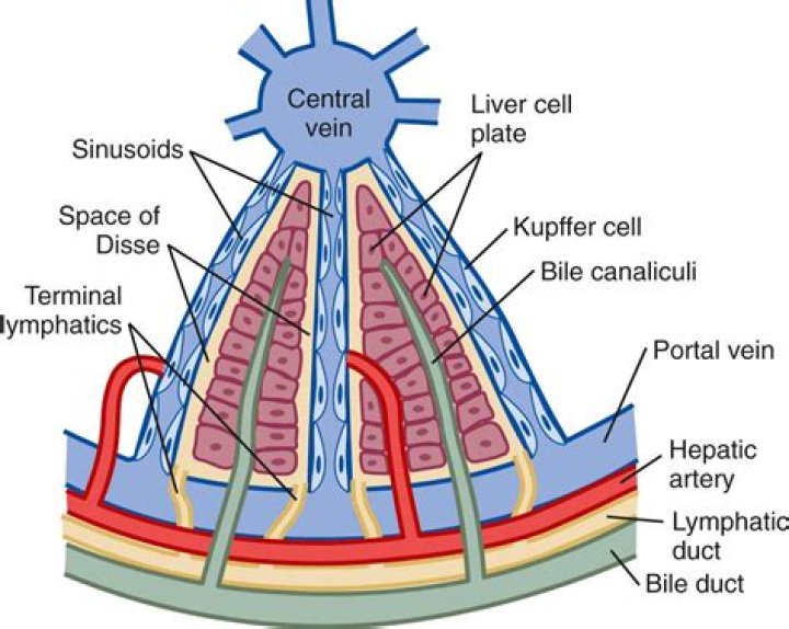
The Disse space lies between hepatocytes and the sinusoids and is also referred to as the perisinusoidal space. The Disse space cannot be visualized on an H&E slide from a normal liver but is visible as a narrow tissue space in electron micrographs (see Fig. 1.11).
Where is Disse?
liver
The perisinusoidal space (or space of Disse) is a location in the liver between a hepatocyte and a sinusoid. It contains the blood plasma….
| Perisinusoidal space | |
|---|---|
| TH | H3.04.05.0.00012 |
| Anatomical terms of microanatomy |
What is the epithelial lining of the liver?
The liver contains two types of epithelial cells, namely, hepatocytes and cholangiocytes. They split from hepatoblasts (embryonic liver stem cells) in mid-gestation and differentiate into structurally and functionally mature cells.
What is perisinusoidal space?
The perisinusoidal space (or space of Disse) is a location in the liver between a hepatocyte and a sinusoid. It contains the blood plasma. Microvilli of hepatocytes extend into this space, allowing proteins and other plasma components from the sinusoids to be absorbed by the hepatocytes.
What is a sinusoid in liver?
Sinusoids are low pressure vascular channels that receive blood from terminal branches of the hepatic artery and portal vein at the periphery of lobules and deliver it into central veins. Sinusoids are lined with endothelial cells and flanked by plates of hepatocytes.
What is a sinusoid anatomy?
sinusoid, irregular tubular space for the passage of blood, taking the place of capillaries and venules in the liver, spleen, and bone marrow. The sinusoids form from branches of the portal vein in the liver and from arterioles (minute arteries) in other organs.
Are Kupffer cells in the space of Disse?
The liver is a complex three-dimensional structure consisting of parenchymal (hepatocytes) and non-parenchymal cells including HSCs, which reside in the perisinusoidal space of Disse, Kupffer cells (KCs), which reside within sinusoids, and endothelial cells, which form the sinusoidal lining.
What is liver sinusoid?
A liver sinusoid is a type of capillary known as a sinusoidal capillary, discontinuous capillary or sinusoid, that is similar to a fenestrated capillary, having discontinuous endothelium that serves as a location for mixing of the oxygen-rich blood from the hepatic artery and the nutrient-rich blood from the portal …
What is the space of Disse?
The Space of Disse, D, located between the sinusoid endothelial cells (S) and the hepatocytes (on the left of the micrograph), is continuous with the sinusoidal lumen. Numerous irregular microvilli, Mv, extend from the hepatocyte surface into the space of Disse, increasing the surface area for metabolic exchange.
What do liver cells look like?
The cells are polygonal in shape and their sides can be in contact either with sinusoids (sinusoidal face) or neighboring hepatocytes (lateral faces). A portion of the lateral faces of hepatocytes is modified to form bile canaliculi.
What is a liver sinusoid?
What is the function of the liver sinusoid?
What is the Disse Space on a normal liver?
The Disse space cannot be visualized on an H&E slide from a normal liver but is visible as a narrow tissue space in electron micrographs (see Fig. 1.11 ). The Disse space contains stellate cells, also called Ito cells or lipocytes; these cells contain lipids and are involved in vitamin A metabolism.
How many liver anatomy stock photos are available on Shutterstock?
Shutterstock’s safe search will exclude restricted content from your search results liver anatomy images 54,607 liver anatomy stock photos, vectors, and illustrations are available royalty-free. See liver anatomy stock video clips
What is microscopic anatomy of the liver?
Microscopic Anatomy of the Liver. Murli Krishna, M.D. The liver is a complex three-dimensional structure that. consists of epithelial and mesenchymal elements arranged in. repetitive microscopic units.