ECG Changes during Myocardial Infarction (MI)
| Location of MI | Leads Affected | Vessel Involved |
|---|---|---|
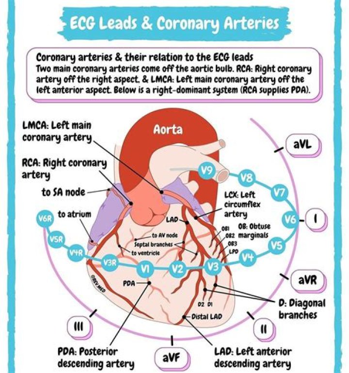
| Inferior wall | II, III, aVF | Right Coronary Artery (RCA) – Posterior descending branch |
| Posterior wall | V1 to V4 | Left Coronary Artery (LCA) – Circumflex branch Right Coronary Artery (RCA) – Posterior descending branch |
Which ECG leads look at septal?
The septum is represented on the ECG by leads V1 and V2, whereas the lateral wall is represented by leads V5, V6, lead I and lead aVL. To make things more complicated, sometimes the LAD “wraps around” the cardiac apex, which is a common anatomic variant.
Can a 12-lead ECG diagnose coronary artery disease?
Resting conventional 12-lead ECG has low sensitivity for detection of coronary artery disease (CAD) and left ventricular hypertrophy (LVH) and low positive predictive value (PPV) for prediction of left ventricular systolic dysfunction (LVSD).
Which leads on a 12-lead ECG are the chest or precordial leads?
For a routine analysis of the heart’s electrical activity an ECG recorded from 12 separate leads is used. A 12-lead ECG consists of three bipolar limb leads (I, II, and III), the unipolar limb leads (AVR, AVL, and AVF), and six unipolar chest leads, also called precordial or V leads, ( , , , , , and ).
What is V4 V5 V6 in ECG?
The electrical activity on an ECG (EKG). The areas represented on the ECG are summarized below: V1, V2 = RV. V3, V4 = septum. V5, V6 = L side of the heart.
Why it is called 12 lead ECG?
The 12-lead ECG displays, as the name implies, 12 leads which are derived by means of 10 electrodes. Three of these leads are easy to understand, since they are simply the result of comparing electrical potentials recorded by two electrodes; one electrode is exploring, while the other is a reference electrode.
Which ECG leads are posterior?
ST elevation in the posterior leads of a posterior ECG (leads V7-V9). Suspicion for a posterior MI must remain high, especially if inferior ST segment elevation is also present.
What is the difference between 5 lead and 12-lead ECG?
5-lead monitoring, which uses 5 electrodes on the torso; and. 12-lead monitoring, which uses 10 electrodes on the torso and limbs.
What are chest leads in ECG?
The chest (precordial) leads (V1, V2, V3, V4, V5 and V6) have the exploring electrodes located anteriorly on the chest wall and the reference point located inside the chest. Hence, the chest leads are excellent for detecting vectors traveling in the horizontal plane.
What is ECG 12 lead?
A 12-lead electrocardiogram (ECG) is a medical test that is recorded using leads, or nodes, attached to the body. Electrocardiograms, sometimes referred to as ECGs, capture the electrical activity of the heart and transfer it to graphed paper.